A poultry post-mortem (necropsy) is a simple, systematic examination of a dead bird's body and internal organs to work out what killed it — and on a Ghanaian farm, done safely and promptly, it is one of the fastest, cheapest ways to turn "my birds are dying and I don't know why" into an actual answer. You do not need to be a vet to open a bird and recognise a few classic patterns: bloody caeca point to coccidiosis, a swollen bursa near the vent points to Gumboro, cheesy patches in the windpipe suggest Newcastle, and a pale, mottled liver hints at aflatoxin or bacterial disease. Picture the common scenario: a keeper is losing three or four growers a day, has already "treated for everything" at the drug shop, and is watching money drain away — when a ten-minute post-mortem on two freshly dead birds would have shown the real problem and saved the rest of the flock. This guide explains why and how to do a basic on-farm post-mortem safely, what the main organs and lesions tell you, and — just as important — when to stop and send the birds to a laboratory or vet.
Written by the Agro Prime Ghana poultry team and reviewed against current veterinary and extension references (MSD Veterinary Manual and university poultry-health extension). Last updated September 2026.
Key takeaways
A basic post-mortem lets you read a dead bird's organs for clues to the cause of death and act on the rest of the flock quickly. Use fresh birds only — a carcass dead more than a few hours, especially in Ghana's heat, decomposes and becomes useless, so necropsy within a couple of hours or refrigerate and do it within a day. Work safely: wear gloves and a mask, wet the feathers with disinfectant, and dispose of the carcass and disinfect tools and surfaces afterwards, because some poultry diseases infect people. Examine organs systematically — trachea, air sacs, lungs, heart, liver, spleen, proventriculus and gizzard, intestines and caeca, kidneys, and the bursa of Fabricius — and match lesions to disease patterns (bloody caeca = coccidiosis; swollen bursa = Gumboro; enlarged leg nerve = Marek's; pale mottled liver = aflatoxin/bacterial). A post-mortem narrows the diagnosis; for a confirmed answer, or anything that could be Newcastle or bird flu, send fresh birds to a veterinary diagnostic laboratory.
Why do a post-mortem on your own farm?
The single biggest reason to learn a basic post-mortem is speed. When birds start dying, the money you lose is not just the dead ones — it is the growth, feed and future birds you lose while you guess. A guess-and-treat approach (buy an antibiotic, a dewormer, a "cure-all" and hope) wastes time and money and often treats the wrong thing entirely: antibiotics do nothing for coccidiosis or a virus, and a dewormer does nothing for aflatoxin. Opening two or three freshly dead or culled sick birds gives you, within minutes, a strong steer on which family of problems you face — gut, respiratory, systemic infection, poison or nutrition — so you can act correctly on the whole flock. Over time it also builds judgement: a keeper who regularly looks inside dead birds learns what "normal" is and spots trouble early. The post-mortem does not replace a vet or a laboratory; it is the fast triage step that tells you what you are probably facing and whether to escalate.
Safety first: protect yourself and the rest of the flock
Before you cut anything, respect two real risks: some poultry diseases can infect people (they are zoonotic), and a careless post-mortem can spread infection around your farm. University poultry-health guidance sets out the basics: work on a flat, hard surface in a well-lit area; wear gloves and, ideally, a face mask; and wet the feathers down with disinfectant first, both to hold down loose feathers and to reduce the aerosols and dust that carry pathogens. Keep water, soap and paper towels within reach, and when you finish, dispose of the carcass and waste properly and disinfect the surface and tools — never work on the litter where your live birds walk, or leave the opened carcass out for other birds, dogs or flies. One point matters enormously in Ghana: if there is any chance you are looking at highly pathogenic avian influenza (bird flu) or an unusual mass die-off, do not open the birds yourself — stop, keep people away, and call your veterinary officer, because that is both a human-health risk and a notifiable disease. Everyday farm biosecurity applies just as much to dead birds as to live ones.
Use only fresh birds — Ghana's heat is your enemy here
The most common mistake is examining a bird that has been dead too long. Once a bird dies its body begins to decompose, and that breakdown creates changes in the organs — a green, softening liver, gassy guts, discoloured tissues — that can be mistaken for real disease lesions, so you end up "diagnosing" rot instead of disease. Extension guidance is blunt that birds dead more than a few hours are not recommended as diagnostic specimens, and in Ghana's heat that window is even shorter. So examine a bird within a couple of hours of death; better still, sacrifice a couple of sick birds showing the flock's typical signs, since a freshly culled bird gives far clearer lesions than one found dead and bloated in the morning. If you cannot do it straight away, refrigerate the carcass (never freeze it, which ruptures cells and ruins the tissue) and examine it within a day.
What you need and how to open the bird
The equipment is deliberately simple: a sharp knife or stout scissors, gloves, a mask, a bowl of disinfectant, and good light. Wet the bird down, lay it on its back, and work through it in the same order every time. The standard approach is to cut through the skin along the inner thighs so the legs fold flat and the bird lies stable, open the skin from vent to neck and reflect it back, then cut through the body wall and remove the breastplate (keel) to open the chest and abdomen. From there you examine, in turn: the mouth, throat and windpipe (trachea); the air sacs; the lungs and heart; the liver, spleen and gall bladder; the proventriculus and gizzard (the two stomachs); the full length of the intestines and the two caeca; the kidneys against the backbone; and the bursa of Fabricius, a small pouch just above the vent that is central to diagnosing Gumboro. Check the sciatic nerves in the thighs and the joints and keel bone too. The skill is not the cutting — it is knowing what each organ should normally look like, so anything abnormal jumps out.
Field note: your phone is part of the toolkit
One habit we strongly recommend is to photograph what you find. Take clear, well-lit pictures of any abnormal organ — the caeca, the bursa, the liver, an enlarged nerve — before the tissue dries out. Those photos let you send exactly what you are seeing to a vet or extension officer on WhatsApp for a second opinion, and build your own reference library of what healthy and diseased organs look like on your farm. A picture of a swollen bursa or a bloody caecum is worth far more to a busy vet than a description down the phone, and it costs nothing.
Reading the organs: what normal and abnormal look like
The heart of a post-mortem is comparison: you are always asking "does this look normal?" The table below sets out what the key structures should look like and what deviations commonly mean, drawing on the MSD Veterinary Manual and university poultry-health guidance. (Our guides to coccidiosis and Gumboro show two of these lesions in detail.)
| Where you look | Normal appearance | Abnormal — and what it suggests |
|---|---|---|
| Windpipe (trachea) | Clear, free of excess mucus | Redness, mucus, blood or cheesy plugs → Newcastle, infectious bronchitis, mycoplasma |
| Air sacs | Thin, clear, shiny — like soap bubbles | Thick, cloudy, yellow-cheesy → airsacculitis (E. coli/mycoplasma, often after a virus) |
| Liver | Firm, even colour (green near gall bladder is normal) | Pale, mottled, fatty → aflatoxin or bacterial disease; white spots → fowl typhoid, blackhead |
| Intestines & caeca | Even walls, no blood or worms | Blood/ballooned caeca → coccidiosis; thick, foul gut → necrotic enteritis |
| Bursa of Fabricius | Cream, fine parallel wrinkles | Swollen, red or bleeding (later shrunken) → Gumboro (infectious bursal disease) |
| Sciatic nerve | Thin, striped, same on both legs | One leg's nerve enlarged/smooth → Marek's disease |
| Keel & joints | Straight keel; shiny white joints | Rubbery keel → rickets; swollen, cloudy joints → infectious arthritis |
Field note: look at the whole picture, not one organ
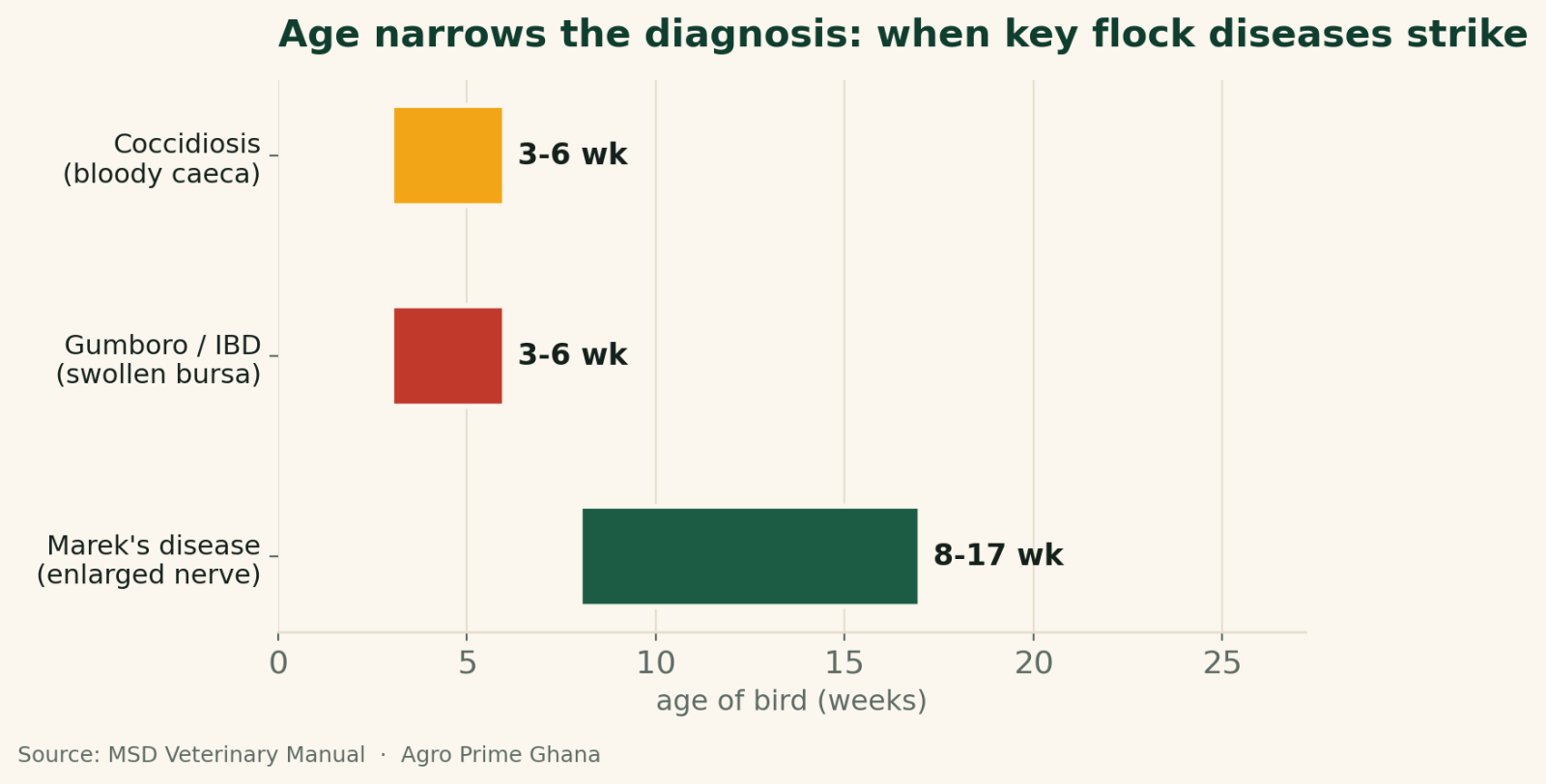
The mistake we most often see is a keeper fixing on one finding and stopping — "the liver has spots, it must be typhoid" — without checking the rest of the bird or the pattern in the flock. A single lesion rarely tells the whole story. We always ask three things together: what age are the birds, what did they do before they died (gasping? bloody droppings? sudden death? slow wasting?), and what do several birds show, not just one. Coccidiosis and Gumboro both hit birds around three to six weeks, but coccidiosis shows bloody caeca while Gumboro shows a swollen bursa — the age narrows it, the lesion confirms it. Open two or three birds, not one, and read the whole animal against the flock's history before you decide.
Matching age and signs to the likely disease
A post-mortem is far more powerful when you combine the lesions with the bird's age, because many diseases have a typical age window (see the chart above). The MSD Veterinary Manual notes that both coccidiosis and Gumboro hit hardest at about 3–6 weeks of age, so a grower dying in that window with bloody caeca is likely coccidiosis while the same-age bird with a swollen, bleeding bursa is likely Gumboro; Marek's disease shows up later, usually in birds 8–17 weeks old, as an enlarged leg nerve and grey tumours. Newcastle strikes at any age with tracheal mucus and cheesy patches, and aflatoxin shows pale, damaged livers in an underperforming flock. Age is one of the first things a good diagnostician asks, because it cuts the list of suspects in half before you even open the bird.
Not sure what you're looking at? Send fresh birds — or ask Agro Prime
A farm post-mortem is triage, not a final verdict. If you cannot read the lesions, if deaths are mounting, or if there is any chance of Newcastle disease or bird flu, get a professional diagnosis: submit two or three freshly dead or culled sick birds, kept cool, to a veterinary diagnostic laboratory or your district veterinary officer, with the flock's age, history and vaccination record. Agro Prime Ghana works with poultry keepers across the country and can help you interpret what you are seeing, point you to the right diagnostic support, and — once you know the problem — supply healthy replacement chicks plus the feed and advice to rebuild the flock. Message us on WhatsApp at +233 24 915 1503 or book a visit.
When to stop and send to a laboratory or vet
Knowing the limits of a farm post-mortem is as important as doing one. Stop and escalate to a veterinary diagnostic laboratory or veterinary officer when the lesions are confusing, when deaths are climbing quickly or across several ages, when the flock is not responding to your field treatment, when you need a confirmed answer for insurance, contracts or a valuable breeding flock, or when you suspect a notifiable disease such as Newcastle or, especially, avian influenza (sudden heavy die-off, purple combs, swollen heads, birds dead with no warning) — in which case you should not open the birds yourself at all. A laboratory can do what your eyes cannot — culture bacteria, test for viruses, examine tissues under a microscope and check for aflatoxin. Submit fresh, cool (not frozen) birds, ideally two or three showing the flock's typical signs, with the full history: age, breed, vaccination record, feed, what you have already given, and the numbers sick and dead. Good farm records make that submission far more useful and the diagnosis far faster.
Disposal and cleaning up afterwards
How you handle the carcass afterwards is part of protecting the rest of your farm. Dead birds and post-mortem waste are a reservoir of whatever killed the bird, so they must be removed from where your live flock lives and disposed of without spreading disease or attracting scavengers, flies and rodents. The safe options are deep burial away from water and the poultry house, or composting or incineration where permitted; do not dump carcasses in the open, feed them to other animals, or leave them near the flock. Disinfect the surface and your tools, wash your hands with soap, and change or clean any clothing and footwear that touched the carcass before returning to live birds. Treating disposal as seriously as the examination is what keeps a diagnostic post-mortem from becoming the source of the next outbreak.
The bottom line on poultry post-mortems
A basic post-mortem is one of the most useful skills a serious poultry keeper can learn: for the price of a sharp knife and ten careful minutes, it turns unexplained deaths into a working diagnosis and lets you act before more birds die. The rules are simple — use only fresh birds (Ghana's heat gives you only a couple of hours), protect yourself with gloves, a mask and disinfectant, examine every organ in the same order every time, and read the lesions together with the birds' age and the flock's history rather than fixing on one finding. Bloody caeca, a swollen bursa, an enlarged leg nerve, a cheesy windpipe, a pale mottled liver — each tells a story once you know what normal looks like. And know your limits: when the picture is unclear, when deaths are mounting, or when there is any hint of Newcastle disease or bird flu, stop, keep people safe, and send fresh birds to a laboratory or call your veterinary officer. Used this way, the humble farm post-mortem becomes your fastest early-warning system.
Frequently asked questions
What is a poultry post-mortem (necropsy)?
Can I do a chicken post-mortem myself without a vet?
How fresh does the bird need to be for a post-mortem?
Is it safe to open a dead chicken? What precautions do I take?
What do bloody caeca or intestines mean in a post-mortem?
How do I tell coccidiosis from Gumboro at post-mortem?
When should I send birds to a laboratory instead?
How should I dispose of the carcass after a post-mortem?
Sources & further reading
- University poultry-health extension — Poultry Necropsy Basics (safety and disinfection, specimen freshness, organ-by-organ examination and normal appearances).
- MSD Veterinary Manual — Infectious Bursal Disease (Gumboro) and Marek's Disease (age of susceptibility and hallmark lesions — swollen bursa, enlarged peripheral nerves).
- Agro Prime Ghana guides: poultry biosecurity, coccidiosis in poultry, Gumboro (infectious bursal disease), and farm record keeping.
About Agro Prime Ghana
Agro Prime Ghana Limited runs a poultry hatchery and mixed agribusiness in Kasoa, supplying day-old chicks and supporting poultry keepers nationwide. These guides combine hands-on hatchery and farm experience with current veterinary references. This article is general educational information, not a substitute for diagnosis by a qualified veterinary or laboratory professional — if your flock is dying, get fresh birds examined and involve your veterinary officer, especially where a notifiable disease is possible. Need healthy replacement chicks, feed or farm support? Book a visit or message us on WhatsApp (+233 24 915 1503).